| "The bars appear on the bearing surface of the foot as convergent ridges which are fused with the sole and united with the frog. By not completing the circle of the wall they allow for expansion of the foot and being part of the wall take weight and provide extra bearing surface and strength at the heels"." |
| Farriery - A Complete Illustrated Guide" John Hickman |
I have to admit that up until recently I had not thought that much about the bars in horses‘ feet. This may have been because no one else seems to be interested in them. In Sisson and Grossman "Anatomy of the Domestic Animals” the bars manage two lines and in Adams"Lameness in Horses there are six. A recent article on "Foot Form and Function" didn‘t even mention them at all.
It was following a weekend course given by Dr Hiltrud Strasser that made me pay more attention to them. She suggested that the bars should be trimmed to allow proper foot function and that prominent bars could be involved in producing heel and "navicular" pain, and this is what made me start to investigate them.
So many authors advise that the bars should not be trimmed, although more recently
I note that some are now suggesting that they should be trimmed to some extent.
It is easy to visualise the bars on the solar surface of the foot, in fact it is also relatively easy to see their position inside the foot by removing the bone, but what is more difficult to visualise is their relationship to the internal structures of the foot.
The atlas of Clinical Anatomy and Comparative Imaging of The Equine Distal Limb (Jean-Marie Denoix) gives further indication of the relationship of the bars in the foot with parasagital, transverse and frontal sectioning of the foot but it is still difficult to visualise them.
I cut out the frog from a number of feet to demonstrate the position, extent and relationship of the bars in the feet, and by doing this I was able to see how close the deep flexor tendon and the navicular bone were to the bars.
Obviously in my investigations I was also considering what the function of the bar was. As stated in Hickman‘s definition above, they take weight and provide extra bearing surface and strength at the heels. They also have a role as part of the anti-slipping and anti-concussion mechanisms of the foot.
If they were absent and the hoof wall ended at the heels, when weight was applied to the leg and foot the walls would expand and the weight would have to be taken by the sole the frog and the digital cushion.
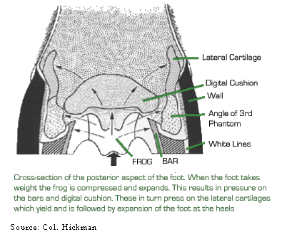
In Col. Hickman‘s Farriery book he includes a figure of the cross-section of the posterior aspect of the foot. The note under it states that when the foot takes weight the frog is compressed and expands. He then suggests that this results in pressure on the bars and the digital cushion, which in turn press on the lateral cartilages which yield and is followed by expansion of the foot at the heels.
I do not agree with this version of events. Any upward forces are in response
to the downward force of the foot when weight is taken on it. The weight has
to be applied to the foot down through the bone column so that it is the pedal
bone that must take the greatest downward force. The reactionary ground force
will initially be greatest at the heel, as the body moves over the foot it will
then transfer forward until it is greatest at the toe prior to the foot being
lifted off the ground.
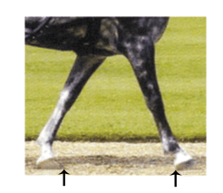
We know this because a shorter forward stride is taken when there is pain at the heel or will have a longer anterior phase and shorter posterior phase of the stride if there is pain at the toe.
So the weight is applied initially through the bone column to the pedal bone. As more weight is applied, because the bones are not in vertical alignment the fetlock and the pastern will drop towards the ground with flexion of the coffin joint and the navicular bone and the deep flexor tendon underneath it will be pressed down on the tissues at the heel.
The downward forces are in effect commencing from the centre of the foot, initially onto the pedal bone and working posteriorly as the pastern drops, and these are being opposed by the reactionary upward ground forces which start from the heel and move anteriorly as full weight is taken.
The downward forces will cause compression of the digital cushion and it is the spreading of these forces to the bars, frog and lateral cartilages that result.

In my opinion the downward movement of the coffin joint, navicular bone and the deep flexor tendon will be onto the bars which, because of their angle, will try to escape by moving downwards and outwards and as they do this, the frog will be compressed. So the effect of the downward pressure and reactionary ground forces is to push the bars outward, the frog and digital cushion to be compressed and apply pressure to the lateral cartilages, further expanding the heels.
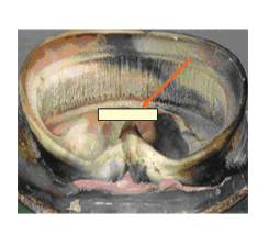
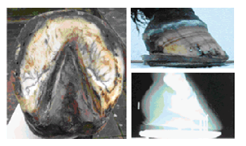
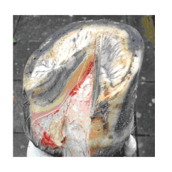
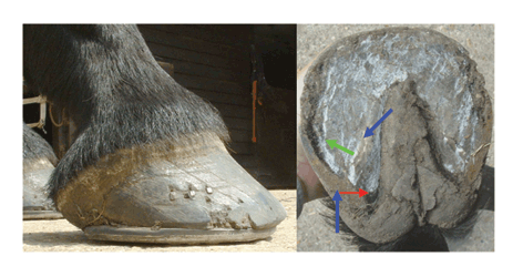
The photographs of the two front feet from a horse . The
left foot shows the bars inside the intact hoof and the right shows the solar
surface and exposure of the bars by removal of the frog.
Left – Shows the extent of the laminae of the bars, extending more than
half way along the frog to the highest point inside the foot. This is continued
to the point of the frog as ridge of sole.
The laminae of the heel and bar make the shape of a "V".
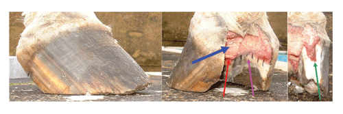
Right – Shows the foot following removal of the shoe and then with the
sole cleaned up. The third and fourth picture are following the removal of
the frog.
In these the fascia of the deep flexor tendon has been exposed and in the
fourth the deep flexor tendon has been cut to expose the navicular bone.
So comparing the planter view of the right fore and the vertical view of the
left fore, the navicular bone will lie just in front of the highest point
inside the foot.
So in what circumstances might they be significant?
Let us consider three types of foot.
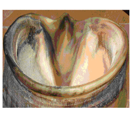
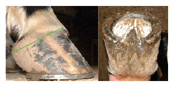
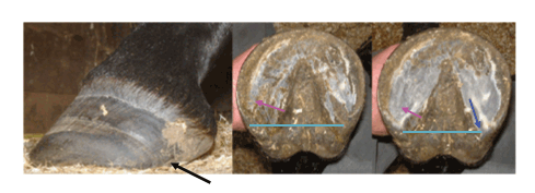
1. The upright foot with high heels.
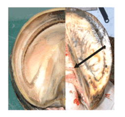
The frog has been cut away and shows the deep flexor tendon which has been cut to expose the navicular bone and also shows the extent of the bars
When the weight is applied to the foot and the pastern drops, the deep flexor tendon will press down onto the bars. Because of their size and the amount of sole between the bars and the hoof wall, they are unable to escape laterally.
These bars will definitely give support to the foot but, because of their rigidity, they won‘t assist in countering concussion. Is it unreasonable to think that rigid bars like this might, in some circumstances, play a part in "navicular" pain.
2. The foot with the curved coronet.
I believe that feet with substantial bars (and soles) do not allow normal expansion of the foot, so that when weight is applied to the foot the digital cushion is compressed, but if the heels are unable to move then the only direction the digital cushion can expand to is onto the lateral cartilages and I believe that it is this that causes the distortion of the coronet.
It seems that the difference between 1. and 2. is to do with the thickness and the strength of the horn of the bar and hoof wall.
3. The foot with the long toe and the collapsed heels.
This shape of foot is common in the thoroughbred which has even weaker horn. For some time I wondered which happened first, the long toe or the collapsed heel? I now believe that it is in fact the bars that collapse first.
When weight is taken on the foot then force is applied to the anterior (internally)
and posterior (externally) edges of the bars.
These weak bars, if they have been left untrimmed, are not strong enough for the forces on them and they will start to bend. The effect of this is to make them curve and I believe that as they curve they will cause the heel to turn in and collapse under the foot.
The pressure from the collapsed heel and the curved bars will cause bruising and "corns"
The "relative" lengthening of the toe with the lower hoof angle delays the breakover. The greater forces at the toe will cause the weak hoof to "flare" and this in turn will pull the sole forward and cause it to collapse.
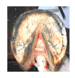
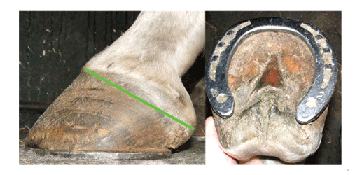
This thoroughbred foot has some collapsing of the heel.
The above picture shows how trimming of the heels will move the posterior part of the ground surface of the hoof back.
The bars have also been trimmed back taking pressure off the seat of corn.
The lateral bar has been trimmed more than the medial side. The lateral bar is now straight and should act as a strut keeping the heel out, against the reactionary ground forces acting on the heel.
I believe that the hoof function of all three types of foot would benefit from trimming of the bars, giving more comfort and sounder horses!
For related articles on the bars please click here
Alternatively download the original document from my downloads page