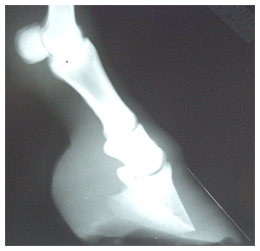
1. To show severity of laminitis:
- Thickness of dorsal wall and sole.
- Separation of P3 from dorsal wall.
- Rotation of P3 - relative to P1 and P2 angle or dorsal hoof wall.
- Sinking of the pedal b style="margin-left:30px;"one.
- Degenerative changes at the tip of the pedal bone.
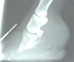
2. To assist in placement of solar support and, in particular, heart-bar shoes.
3. Repeat radiographs over time - to indicate progress of the case.
How should it be carried out?
- Ideally on a flat level surface.
- With the horse standing square.
- Markers applied to:
- The dorsal wall of the hoof up to the hair-line.
- The point of the frog. (I put a drawing pin in 1cm from the point)
- The top edge of the block the horse is standing on.
- The hair line at the heel. (I believe that this needs to be marked if we are to make a measurement of the position of the extensor process of P3 in relation to the hoof.)
- The X-ray beam should be at right angles to and centred level with the estimated position of the mid point of the solar surface of the pedal bone. (There is a large variation on the advice given about the height that the X-ray beam should be, from "1cm distal to the coronary band" to "1cm above the bottom of the hoof". In practice, I put the foot on the block and the X-ray machine on the ground and aim it at right angles to the foot.)
| I recommend the clear and concise “Radiography of the Equine Foot – Techniques for enhancing the quality of your films” R.F.Redden, DVM Equine Podiatry Monograph Series. Published by Nanric Inc. www.nanric.com |
The problems and limitations of radiography and interpretation
Flat Level Surface:
Finding a level flat surface to carry out X-rays may be easy in the hospital situation but virtually impossible in the vast majority of my client‘s yards.
If the X-rays are not taken on a flat surface it is more difficult to measure the sole thickness, the solar angle of P3, and the drop of P3.
Should we be trying to X-ray acute laminitic cases on site, getting less satisfactory radiographs, or should we be transporting them to a hospital to get true diagnostic plates? I think that the benefits of getting better radiographs is far outweighed by the potential worsening of the condition by transporting the animal and I make the best job I can on site.
Standing Square:
The X-ray plate has to extend below the level of the sole to include the whole foot so that, in order to be able to stand square, the horse has to stand on two blocks.
I suspect that many will radiograph the horse with only one foot on a block, which is how I do it, but we have to be aware of the limitations of doing so.
We should get a satisfactory lateral radiograph of the foot but we will not get a true foot pastern axis, and thus rotation of the pedal bone in relation to this.
The Hoof Pastern Axis (HPA)
Even if the horse stands on two blocks do we get a true picture of the hoof pastern axis?
We may find a difference if the horse is taking weight on the foot or not.
We may find a difference if the head is held high or low.
Some people feel we should radiograph the foot when the horse is standing with the cannon bone vertical, suggesting that this will give us the true FPA.
Having taken photographs of the feet of over 400 horses and measured the dorsal wall angle of many more, I disagree with this.
I suggest that the angles of the hoof are remarkably consistent and are the same angles as those of the pedal bone. (See Strassers' Plastic Sheet)
I find the angle of the line of the coronet (from the hair-line at the heel to the hair-line at the toe) to the proximal dorsal wall is very close to 105° . The sum of other two angles, the proximal dorsal wall to solar angle and the solar angle to the line of the coronet will add up to around 75° . (All three angles together must add up to 180° .)
This means that if dorsal wall to ground angle is 45° then the line of the coronet is 30° to the ground. If the dorsal wall is 55° then the coronet will be 20° to the ground.
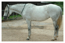
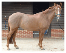
Many, if not most, non-laminitic horses with a 55° dorsal wall angle will stand under if standing square. (Standing with their legs leaning back under the body.) Horses with a dorsal wall angle of 50° or less will stand more commonly with an upright straight leg.
Both of these horses will have a straight, but different, hoof pastern axis.
If a horse has a 55° hoof wall, a straight HPA and stands under, and the heel is trimmed so that the dorsal wall angle is lowered to 50° then the position of the leg, when the horse stands square, will move more upright. It will again have a straight HPA but it will be lower than before.
At a foot course I attended 15 months ago two of the lecturers, one a vet and the other a farrier, both stated that a horse has one hoof pastern axis. When I questioned this and pointed out that a horse could have more than one, the reply I got was that they had one "correct" hoof pastern axis.
Is the "correct" HPA when the hoof is at a higher angle and the horse is standing under, or as is often suggested when the horse stands with a vertical leg?
So I suggest that if we radiograph all horses with their feet positioned so they stand with a vertical cannon then the horse will not be in its normal stance and we will have produced a broken HPA.
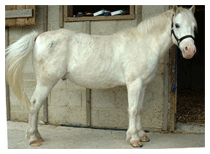
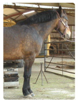
Some horses (particularly ponies) with chronic laminitis, will have high dorsal hoof wall angles (the proximal part if there is deviation of the wall) but instead of standing under, as the non-laminitic horse does, will stand with a vertical leg. I believe that this way they can put more of their weight down on the heel and put less pressure down on the toe.
When radiographed, these ponies will be termed to have "phalangeal rotation". (Realignment of P3 - The basis for treating chronic laminitis S.G.O‘Grady BVSc, MRCVS Paper presented at BEVA day meeting on laminitis Dec 02 also at AAEP annual convention 2003)www.equipodiatry.com
It is these animals with phalangeal rotation that are considered to have excessive pull of the deep flexor tendon and for this reason are considered to be suitable candidates for deep flexor tendon tenotomy. I suggest that, because they stand with a vertical leg when they have an upright foot, they are indicating that the tilt of the pedal bone is causing pain in the foot and that the contracture of the deep flexor tendon is in response to the pain. (See "How should we trim the chronic founder foot?").